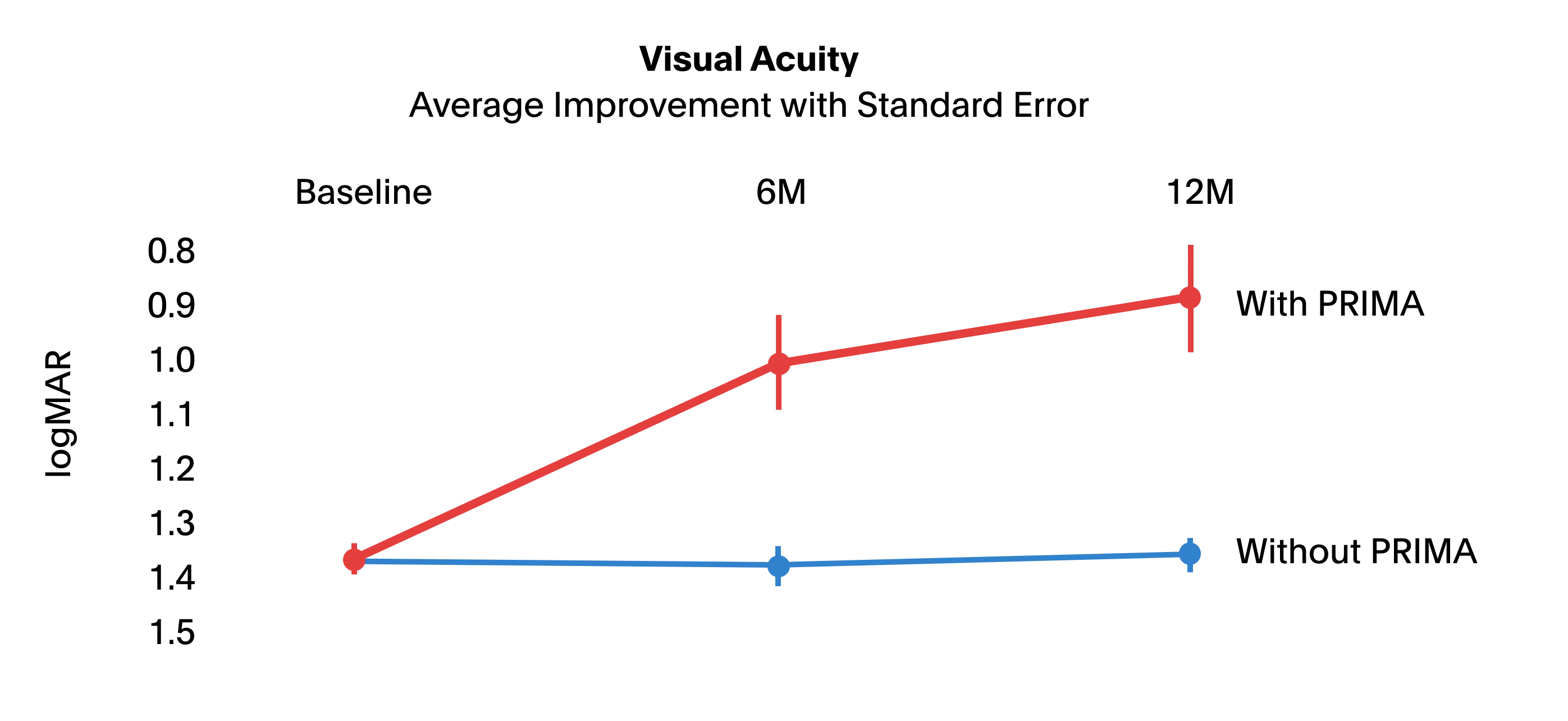
Primary Endpoint at 12 Months
80%
of participants achieved clinically meaningful visual acuity improvement (≥10 ETDRS letters) n=32, 95% CI, 66-94, P<0.0011
Information for Healthcare Professionals
Geographic atrophy is an advanced form of "dry" AMD, characterized by the progressive and irreversible loss of photoreceptors and retinal pigment epithelial cells, culminating in profound central vision impairment.
Current therapeutic options (complement inhibitors and cell therapies) are only partially effective in slowing disease progression and are unable to stop the natural course of the disease. Conventional low-vision aids rely on magnification and do not address the underlying sensory deficit.1
PRIMA bypasses damaged photoreceptors to generate a new central visual percept while preserving natural peripheral vision.
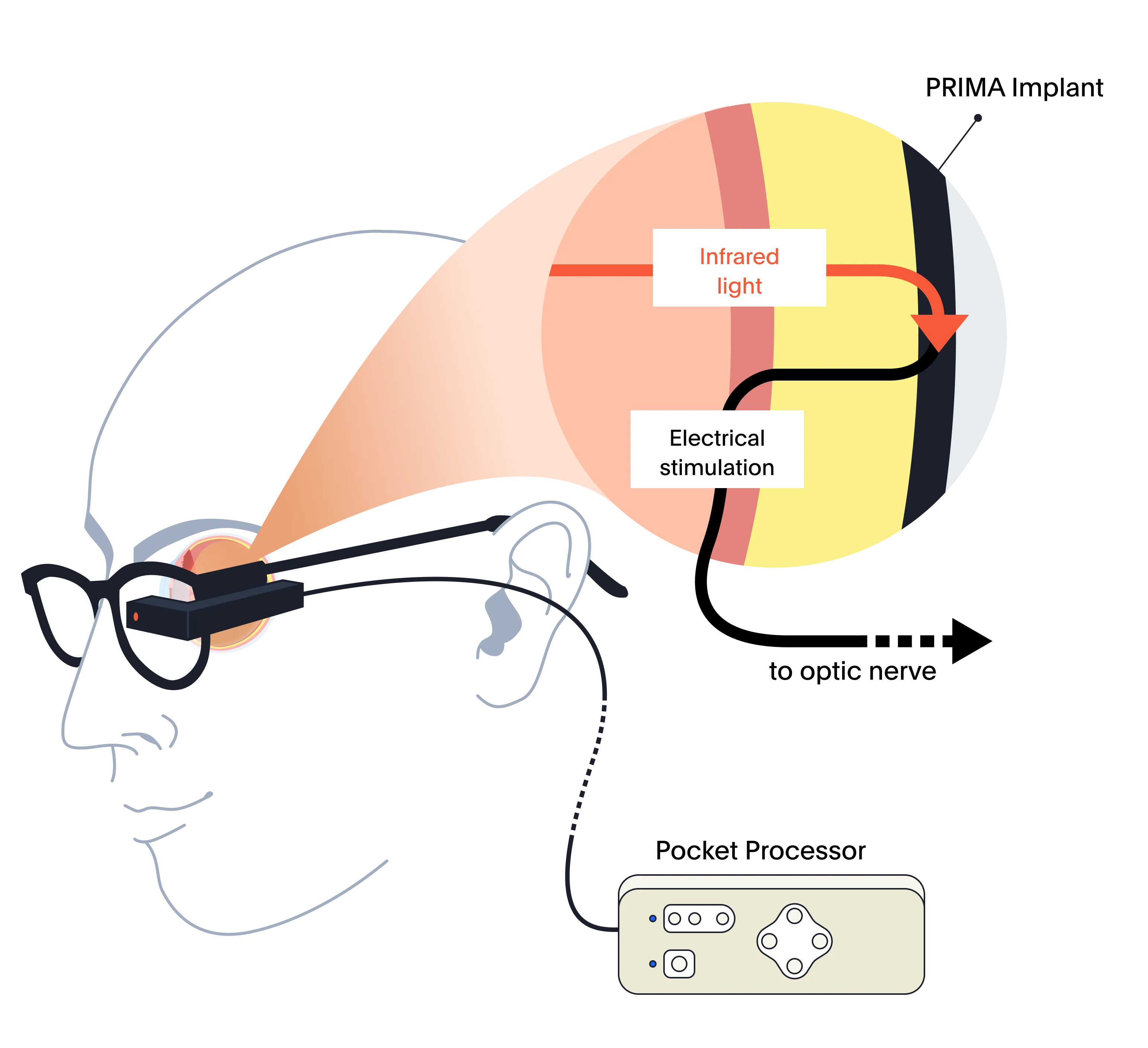
A built-in camera on the glasses captures the visual scene. A pocket processor manages real-time signal processing and wireless transmission of stimulation parameters.
An integrated projector transmits patterned near-infrared light through the pupil onto a photovoltaic array (2 × 2mm; 30µm thick) positioned subretinally.
378 photovoltaic pixels generate localized electrical pulses that stimulate adjacent bipolar cells of the inner nuclear layer. Processed signals traverse the optic nerve and are interpreted in the visual cortex.
The following table outlines some of the key eligibility criteria:
| Criterion | Specification |
|---|---|
| Age | 60 years or older |
| Diagnosis | Bilateral GA due to AMD |
| Visual acuity | BCVA ≥ 1.2 logMAR (≤ 20/320 Snellen; ≤ 0.06 decimal) |
| Lesion size | Foveal atrophy > 4.5mm2, > 2.4mm minimum diameter |
| Rehabilitation capacity | Motivated and able to attend scheduled follow-up visits and complete the vision rehabilitation program |
PRIMA may not be suitable if the patient has certain other eye conditions, active infections, or health conditions that could affect surgery or recovery.
The PRIMA clinical trial was a prospective, multicenter, single-group (single-arm) study, evaluating the safety and efficacy of the PRIMA System in 38 patients with GA involving the central macula due to AMD.
The results were published in the The New England Journal of Medicine in October 2025.1 In a follow-up NEJM editorial, author Jacque Duncan, M.D., described PRIMA as “the first treatment to restore vision” in patients with advanced GA.2
80%
of participants achieved clinically meaningful visual acuity improvement (≥10 ETDRS letters) n=32, 95% CI, 66-94, P<0.0011
~25 letters
at 12 months; best individual improvement: 59 letters1
84%
reported reading letters, numbers, and words with artificial vision1
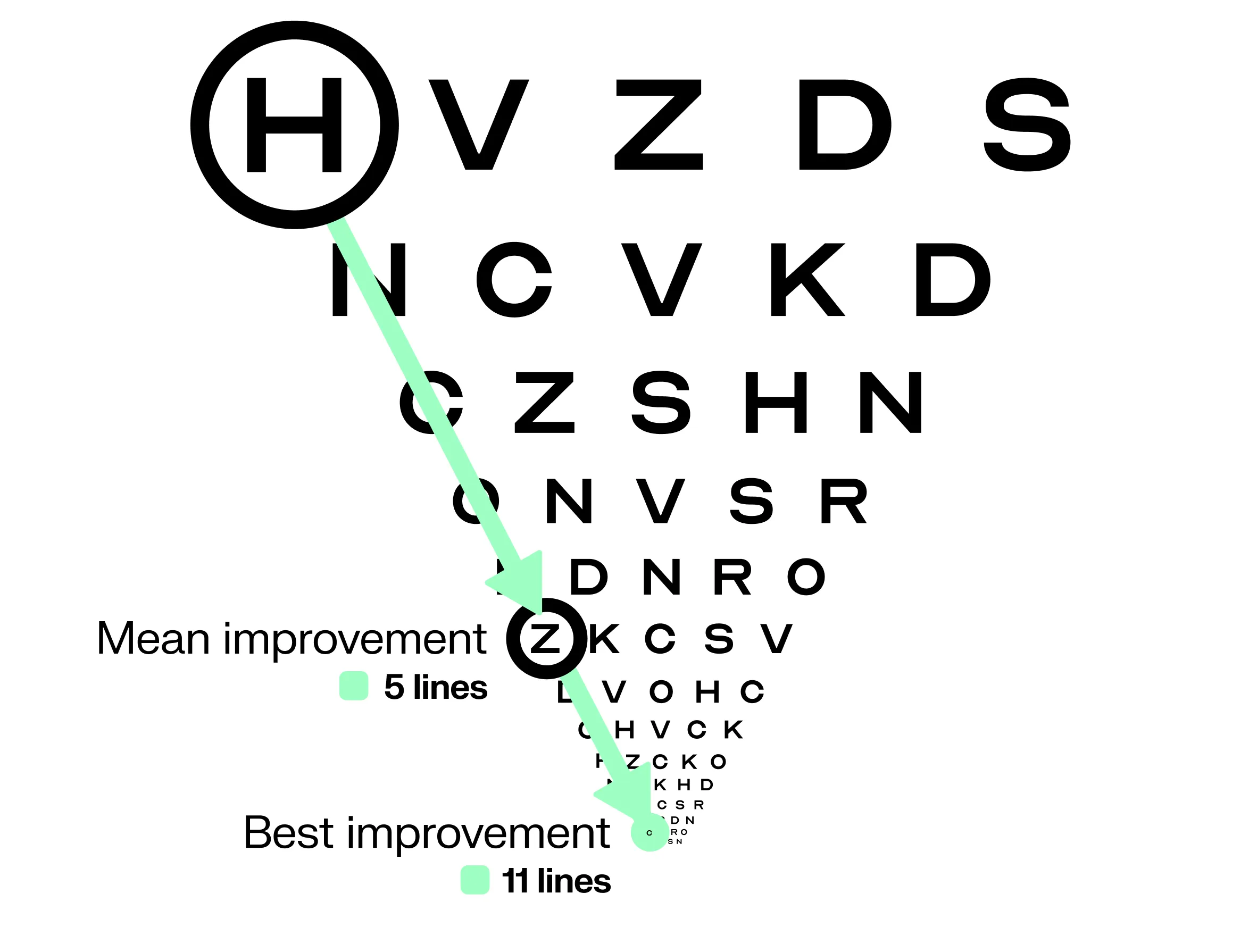
Data collected over a period of 12 months showed that the PRIMA Implant can be safely implanted under the atrophic macula and restore central vision while preserving natural peripheral vision.1
The safety profile of the PRIMA System was evaluated across all 38 participants in the PRIMA clinical study through 12 months of follow-up. The overall profile was consistent with that expected for subretinal vitreoretinal surgery, and the benefit-risk balance was considered favourable by the independent Data Safety Monitoring Board.1
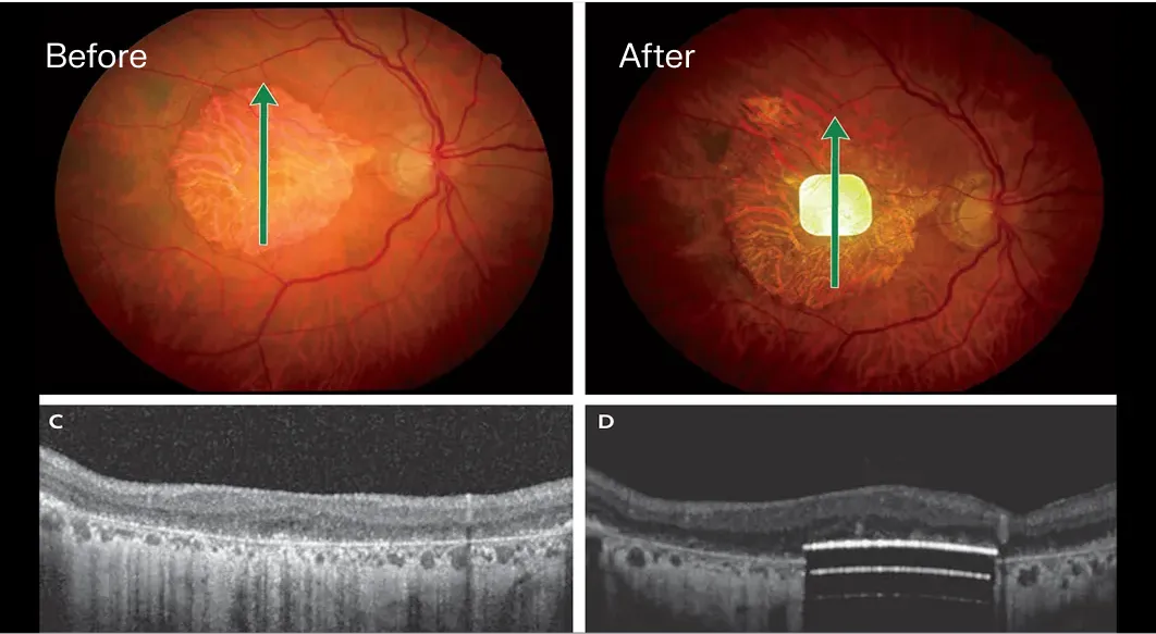
Colour fundus and OCT images of the PRIMA Implant, before and 12 months after implantation.
The implant is delivered subretinally via pars plana vitrectomy. Surgery is an average duration of 1 to 2 hours at a certified PRIMA center, performed by a trained vitreoretinal surgeon.
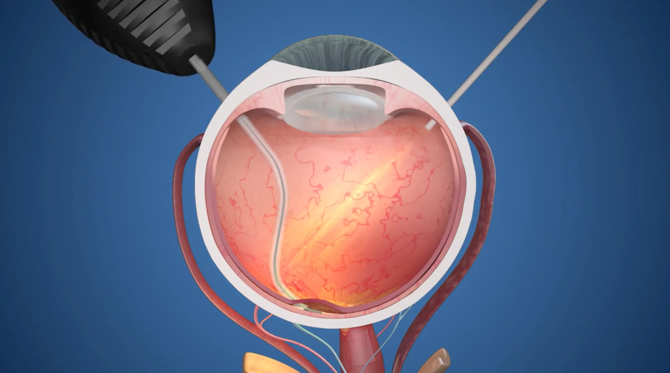
This video shows how the PRIMA Implant is placed beneath the retina, and its mechanism of action.
Post-implantation, patients follow a structured rehabilitation program of 60 hours of training with a dedicated vision rehabilitation specialist. Patients who follow the full rehabilitation program are more likely to achieve the best possible outcome.
The referring retinal specialist remains part of the care team, resuming ongoing follow-up after rehabilitation.
“These patients who had lost their central vision completely can again read letters, numbers, and also even books.”

Professor & Chairman, Dept. of Ophthalmology, University of Bonn
“It's the first time an artificial retinal implant has worked so well in a large study, with a procedure that could be carried out by multiple surgeons.”

Chair, Dept. of Ophthalmology, University of Pittsburgh; Founder, Institut de la Vision, Paris
“PRIMA needed to have a procedure that was something you could easily do in your standard practice.”

Vitreoretinal Surgeon, Moorfields Eye Hospital; Associate Professor, Institute of Ophthalmology, University College London